
Embryo Biopsy and the PGT Lab Process: What Happens After the Sample Is Taken
Embryo biopsy is the laboratory step that lets some IVF cycles test an embryo's chromosomes before transfer. This guide walks through when the biopsy happens, how embryologists sample trophectoderm cells, what the genetics lab does with the sample, and how to read euploid, mosaic, and aneuploid results.
What an embryo biopsy is and why clinics use it
An embryo biopsy is a laboratory step in some in vitro fertilization (IVF) cycles in which a small sample of cells is taken from an embryo so the cells can be tested for chromosome number. In the United States this testing is usually called preimplantation genetic testing for aneuploidy, or PGT-A. The goal is to learn whether an embryo has the expected number of chromosomes before it is transferred, so a clinic and patient can decide which embryo to use first.
It helps to separate the biopsy (the physical sampling of the embryo) from the genetic test itself. The biopsy is done in an embryology lab; the genetic analysis happens afterward in a genetics laboratory. Our companion guide on PGT-A explains what the test looks for and what the evidence shows about outcomes. This article focuses on the procedure: what is done to the embryo, when, and what happens to the sample.
When the biopsy takes place in the cycle
Most biopsies are performed on day 5 or day 6 after fertilization, once the embryo has reached the blastocyst stage. A blastocyst is an embryo that has formed a fluid-filled cavity and two distinct cell types. Reaching this stage matters because the embryo has separated into an inner cell mass that becomes the baby and an outer layer called the trophectoderm that becomes the placenta.
Performing the biopsy at the blastocyst stage is preferred because the trophectoderm provides enough cells to test without disturbing the cells that would form the fetus. Some clinics also offer biopsy at an earlier cleavage stage, but blastocyst-stage biopsy is the more common approach in the United States today. If an embryo does not grow to the blastocyst stage, it generally cannot be biopsied.
How the biopsy is performed
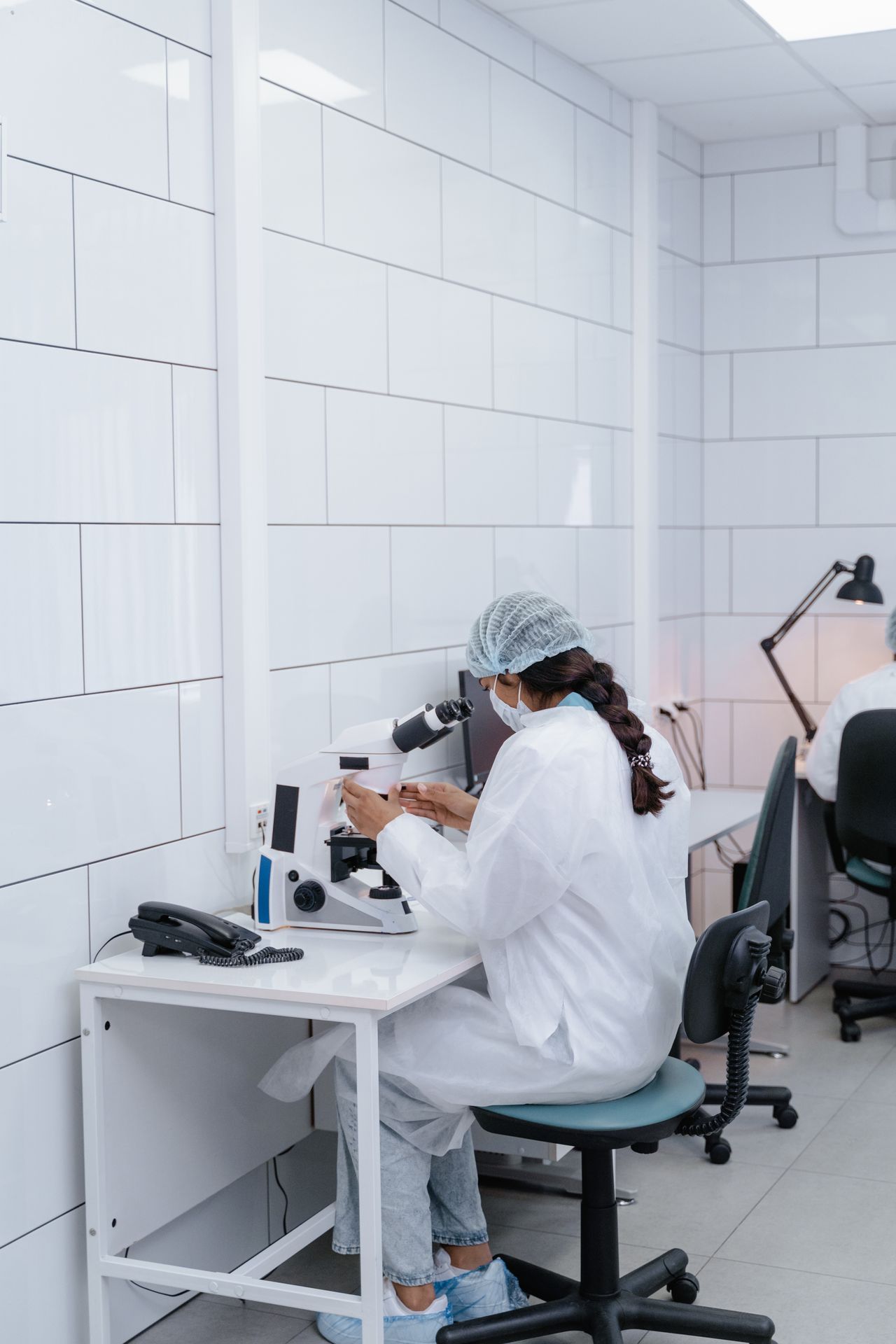
The embryo is held gently in place under a microscope. Using a focused laser, the embryologist makes a small opening in the shell that surrounds the embryo, called the zona pellucida. A few trophectoderm cells are then removed through that opening. Typically between three and ten cells are sampled, depending on the embryo's size and the clinic's protocol.
The removed cells are placed in a small tube and labeled with a unique identifier that links them back to that specific embryo. The embryo itself is returned to the incubator, and in most cases it is frozen, a process called vitrification, while the genetic results are pending. You can read more about frozen embryo transfer to understand what happens after the results return.
What the laboratory does with the sample
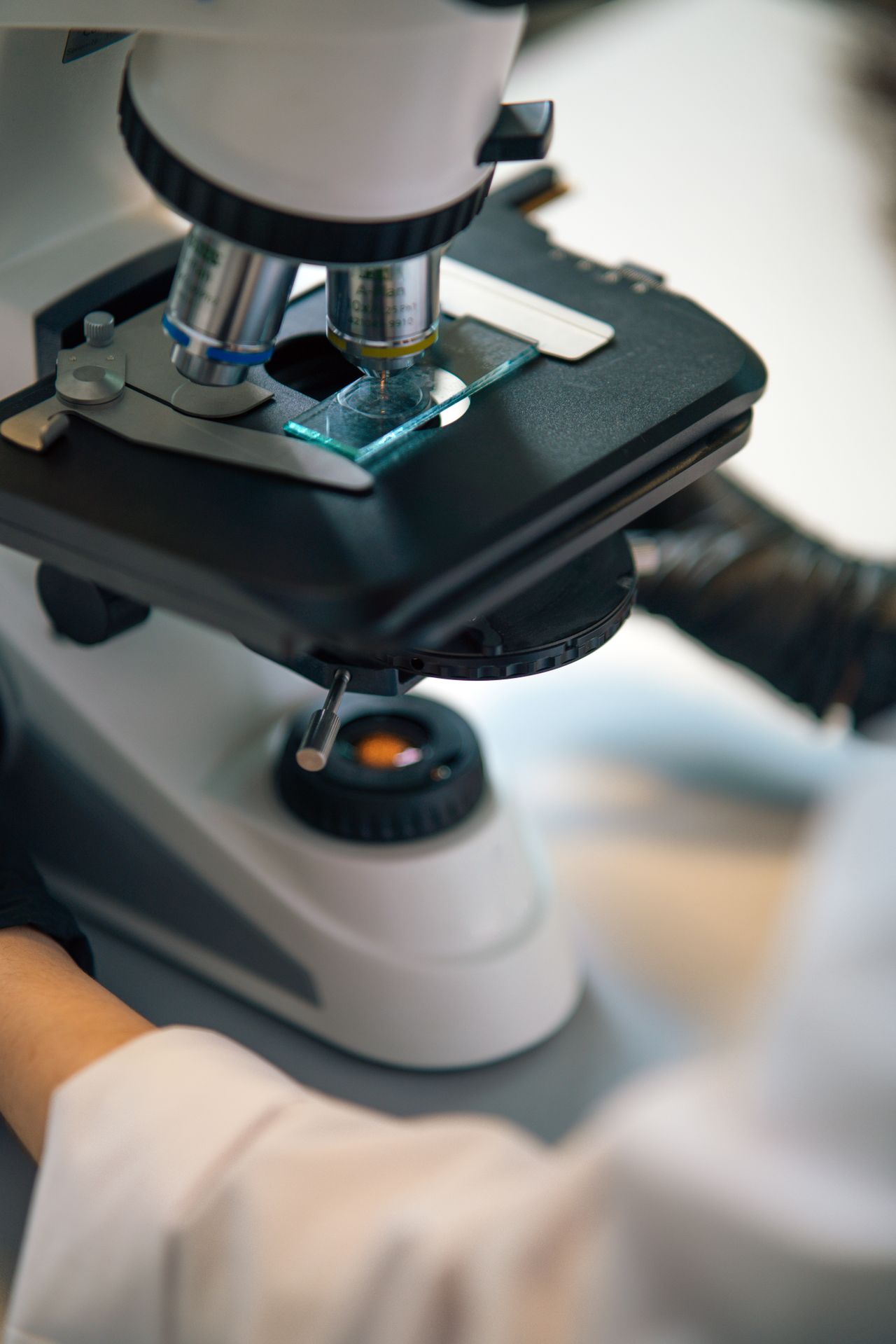
Once the genetics lab receives the sample, the small number of cells means the DNA must first be amplified, because a handful of cells does not contain enough genetic material to analyze directly. After amplification, the lab uses a platform such as next-generation sequencing or a microarray to count the chromosomes across all 23 pairs.
The laboratory then reports the result in categories. A euploid embryo has the expected number of chromosomes. An aneuploid embryo has too many or too few chromosomes for one or more pairs. A mosaic embryo contains a mixture of normal and abnormal cells. Each clinic presents these categories a little differently, and the meaning of a mosaic result in particular depends on the clinic's reporting method and thresholds.
Understanding the results
A euploid result is generally considered favorable for transfer, but it does not predict pregnancy with certainty, and it does not rule out other conditions. Aneuploid embryos are less likely to implant and are more likely to end in miscarriage, and in some cases they correspond to known chromosomal conditions. Mosaic results are interpreted cautiously; many clinics have written policies about whether and how mosaic embryos may be used, and those policies vary.
It is important to remember that PGT-A reports chromosome number only. It does not assess every genetic condition, does not measure embryo quality in a way that replaces morphological grading, and does not tell you whether a pregnancy will be healthy. IVF success rates depend on many factors beyond chromosome testing.
Embryo storage while awaiting results
Because the genetic report usually takes about one to two weeks, the biopsied embryo is vitrified and stored. Annual storage is a separate consideration; our guide to embryo storage costs outlines what to ask about fees and limits. When results are ready, your clinic will discuss which embryo or embryos to transfer and when.
Limits, costs, and questions to ask
PGT-A adds laboratory fees on top of the IVF cycle, and insurance coverage varies by state and plan. The test reduces uncertainty about chromosome number but cannot eliminate it. Inaccurate results, while uncommon, are possible because the sample comes from a small number of cells. Ask your clinic how mosaic results are handled, what platform is used, and how embryos are tracked so the correct sample is matched to the correct embryo.
References
- American Society for Reproductive Medicine (ASRM): patient education and committee opinions — asrm.org
- Society for Assisted Reproductive Technology (SART): clinic-level outcome data — sart.org
- Centers for Disease Control and Prevention (CDC): assisted reproductive technology surveillance — cdc.gov/art
This article is general educational information and is not medical advice. Discuss your specific situation with a licensed medical professional.